Advanced Optical Imaging Suite
The Advanced Optical Imaging Suite within the MNP Group is a cutting-edge facility designed to probe the smallest scales of matter with exceptional precision. Combining world-class imaging technologies with expert support, the suite plays a vital role in advancing interdisciplinary research across physics, chemistry, biology, and materials science.
Our suite is equipped with a range of state-of-the-art optical microscopy platforms, including confocal, super-resolution STED (Stimulated Emission Depletion), fluorescence lifetime imaging microscopy (FLIM), and total internal reflection fluorescence (TIRF) microscopes. These systems enable researchers to visualise molecular structures, dynamic processes, and nanoscale interactions in real time with sub-diffraction resolution and spectral accuracy.
This facility supports a wide variety of applications—from imaging individual proteins and live cells to characterising nanostructured materials and soft matter systems. Whether investigating fundamental biophysical mechanisms or developing next-generation nanotechnologies, the Advanced Optical Imaging Suite empowers researchers to capture data that was once beyond the limits of conventional microscopy.
Situated at the heart of our research labs, the suite is fully integrated with the MNP Group’s wider analytical capabilities, including spectroscopy, nanofabrication, and surface characterisation. It not only accelerates the pace of discovery but also fosters collaboration across disciplines, making it a cornerstone of innovation and training for students, researchers, and industry partners alike.
In addition to its technical capabilities, the suite offers comprehensive training programmes, user inductions, and one-to-one technical support for students, postdoctoral researchers, and academic collaborators. Our dedicated imaging specialists provide guidance on experimental design, data acquisition, and image analysis, ensuring users can maximise the potential of each instrument. From first-time users to experienced microscopists, the suite supports a vibrant, collaborative research environment that promotes scientific excellence and innovation.
Advanced Microscopy Platform
Our suite features a curated selection of cutting-edge microscopes, purpose-built to support high-level research and detailed nanoscale analysis. Each system is chosen not only for its precision and versatility but also to encourage hands-on learning, empower researchers, and drive innovation across disciplines.
Deformation Microscope - Nikon Eclipse Ti Inverted Microscope
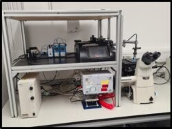 Deformation Microscope system, housed within our advanced microscopy suite, is a cutting-edge platform designed for high-speed imaging and mechanical manipulation of biological samples. At its core is the Nikon Eclipse Ti inverted research microscope, renowned for its stability, modularity, and optical precision.
The system is equipped with the Nikon Intensilight C-HGF-1 fluorescence light source, providing a long-lasting, high-intensity illumination ideal for time-lapse fluorescence imaging with minimal photobleaching. Integrated into the system is the Photron Fastcam SA5 high speed-camera, capable of capturing up to 7,500 frames per second (fps) at full resolution (1024 x 1024 pixels) and significantly higher frame rates at reduced resolutions – making it ideal for studying rapid biomechanical events such as cell deformation, membrane rupture, or cytoskeletal dynamics under stress. This microscope is further enhanced with a Harvard Apparatus (HA) syringe drivers as well as air-based pumping systems from Dolomite and Fluigent, enabling precise, programmable mechanical loading (e.g: stretch, compression, or shear) of live cells, tissues, or synthetic constructs, supporting research in mechanobiology, cell-matrix interactions, tissue engineering, and biomechanics.
The system supports multi-channel fluorescence imaging, with a filter cube turret and compatibility with a wide range of excitation/emission filter sets. Controlled via Nikon NIS-Elements or Photron FASTCAM Viewer software, the platform allows synchronized acquisition of mechanical and optical data, ideal for quantitative analysis of cell mechanics, viscoelastic responses, and mechano-transduction pathways.
This powerful setup makes it a vital resource for interdisciplinary research across biophysics, regenerative medicine, and collaborative projects within the School of Physics and Astronomy and its partner departments.
Photothermal Laser System - Coherent Mira Optima 900-D
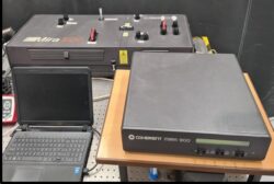 Our Mira 900-D is powerful and versatile Titanium Sapphire (Ti:Sapphire) tunable laser system designed for ultrafast photonics applications. Pumped by a high-power, low-noise Verdi-V10 green diode-pumped solid-state (DPSS) laser, delivers pulses with a duration of less that 115 femtoseconds (fs) in fs mode and less than 2 picoseconds (ps) in ps mode, with a tuning range spanning from 700 – 980 nm. This makes it ideally suited for a range of time-resolved spectroscopic techniques. The system features a seamless mode-switching capability between continuous-wave (CW), ps and fs operations, thanks to an innovative design that incorporates passive kerr lens mode-locking for enhanced stability and ease of use. The integrated Optima control system includes a fast photodiode, relative power monitor, β-lock system, CW detector, humidity sensor and an automatic starter. This suite of diagnostic and feedback components allows precise adjustment and monitoring of output parameters such as pulse width, power and wavelength. This result is a reliable, user-friendly platform for advanced ultrafast laser experimentation.
The photothermal laser setup, built around the Mira 900-D, enables non-invasive, high-sensitivity detection of optical absorption at the nanoscale. By using a modulated laser beam to locally heat the sample, it can detect minute changes in thermal properties, which are captured through temperature changes in the medium of choice.
Key applications of this system include nanoparticle characterization, detection of weakly absorbing substances, and the study of thermally responsive biomaterials. The Mira 900-D thus serves as a critical tool in cutting-edge photothermal research, supporting interdisciplinary investigations across nanotechnology, biophysics, and materials science.
3D Double Helix Super-Resolution / Total Internal Reflection Fluorescence (TIRF) Microscopy
 The 3D Double Helix Super-Resolution/Total Internal Reflection Fluorescence (TIRF) Microscopy system, is a cutting-edge imaging platform enables single-molecule imaging across a variety of modalities. The Double Helix-Point Spread Function (DH-PSF) approach encodes axial depth information into a rotating point spread function, enabling precise 3D localization of single molecules with lateral resolution of ~20-30 nm and axial resolution of ~20-50 nm, over an extended axial range of up to several microns. This is ideal for tracking the motion of proteins in cells or to reveal the nanoscale organisation of proteins and subcellular structures. TIRF microscopy, which restricts fluorescence excitation to a thin region (~100-200 nm) adjacent to the glass-water interface, drastically reduces background signal and photobleaching, making it ideal for imaging near-membrane events in live cells or to perform super-resolution imaging with high spatial 3D resolution.
The system is equipped with a high numerical aperture immersion objectives (water 1.27 NA/Oil 1.49 NA), scientific Complementary Metal-Oxide Semiconductor (sCMOS) cameras for single-molecule detection, and multiband fluorescence filter sets for multi-colour imaging. Laser-based illumination with adjustable TIRF angles allows for controlled evanescent field generation. Additionally, the system includes vibration isolation, a temperature-controlled environmental chamber for live-cell imaging, and optional focus-lock systems to maintain stability during long-term time-lapse experiments. This configuration offers a powerful solution for investigating nanoscale biological processes such as vesicle trafficking, receptor dynamics, focal adhesion behaviour, and cytoskeletal interactions with high spatial and temporal resolution.
Our facility is equipped with cutting-edge hardware and specialized software for drift correction, localization analysis, and 3D reconstruction. The instrument is available to academic users across disciplines including molecular biology, biochemistry, neuroscience, and materials science. Full training and technical support are offered to new users, and our microscopy specialists are available for consultation on experimental design, fluorophore selection, and data interpretation.
Confocal - Renishaw inVia Raman Microscope
 The Renishaw inVia Confocal Raman Microscope, operated through the Windows-based Raman Environment (WiRE) software, is a high-performance spectroscopic instrument designed for advanced chemical imaging and molecular characterization. Covering a spectral range from 200 nm to 2200 nm and supporting multiple laser excitations from 229 nm to 1064 nm, it offers exceptional spectral resolution down to 0.3 cm⁻¹ (FWHM). This state-of-the-art system enables non-destructive analysis with high spatial precision and sensitivity, making it ideal for both materials and life sciences research. The system we have is distinctly different from the normal systems in that; is linked to an inverted confocal Fluorescence microscope system from Leica-DMi8 microscope, SP8 confocal system controlled by Leica LAS-X software.
One of the standout features of the inVia system is its confocal Raman spectroscopy capability, which employs confocal optics to eliminate out-of-focus light. This results in detailed 3D chemical mapping of complex, heterogeneous samples such as nanomaterials, thin films, and live cells. The microscope supports multiple laser wavelengths—typically 532 nm and 785 nm in our system—allowing researchers to tailor analyses based on the sample type, optimize Raman signal quality, and minimize background fluorescence. Its high sensitivity and resolution also make it well-suited for detecting subtle spectral variations, essential for studying polymorphs, mapping stress and strain in materials, and monitoring phase transitions.
Applications of the Renishaw inVia system span a wide range of disciplines. In materials science, it facilitates the characterization of advanced materials such as graphene, carbon nanotubes, semiconductors, polymers, and ceramics. In the life sciences and biochemistry, it enables non-invasive examination of cells, tissues, and biomolecules without the need for fluorescent labelling. The pharmaceutical sector benefits from its ability to map drug distributions, identify polymorphic forms, and assess tablet formulation quality. Additionally, it plays a critical role in forensic and environmental sciences, aiding in the detection of trace substances, pigments, and microplastics.
Located within the School of Physics & Astronomy, the Renishaw inVia Confocal Raman Microscope is a core instrument in the analytical microscopy suite. It supports interdisciplinary research across physics, chemistry, biology, engineering, and materials science. The system is actively used for collaborative projects, postgraduate research, and specialized training, making it an essential resource for advancing scientific discovery.
Reflection Interference Contrast Microscopy (RICM) - Zeiss Microscope with Incubator XLmulti SI
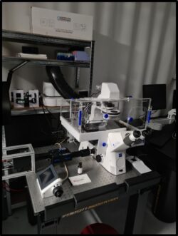 The Advanced Optical Imaging Facility in the School of Physics & Astronomy houses a state-of-the-art RICM system built around a ZEISS inverted research microscope, designed for advanced nanoscale and live-cell imaging. This integrated platform combines high-resolution interference-based techniques with versatile fluorescence and brightfield imaging, providing researchers with a powerful and flexible toolset.
At its core, the system employs Reflection Interference Contrast Microscopy (RICM) to deliver label-free, high-sensitivity imaging of surface contacts, capable of detecting sub-nanometer variations between live cells, lipid vesicles, thin films, and reflective substrates. This enables detailed investigation of cell–substrate adhesion, vesicle–surface interactions, and nanoscale membrane mechanics under physiologically relevant conditions.
The ZEISS microscope enhances RICM with a full suite of imaging modalities, including brightfield, phase contrast, DIC, and fluorescence. Fluorescence imaging is supported by multi-channel LED or laser illumination with a motorized filter turret, accommodating a wide range of dyes (DAPI, FITC, TRITC, Cy5). High-NA objectives (40x/1.30, 63x/1.40), a sensitive sCMOS/CCD camera, motorized stage control, and ZEN software ensure precise, reproducible experiments across multiple applications.
For extended live-cell studies, the integrated PeCon XLmulti S1 incubation system maintains stable temperature (10–50 °C), CO₂, and humidity, ensuring long-term sample viability in multi-well plates, petri dishes, or microfluidic devices. This allows researchers to monitor cellular dynamics such as mitosis, cytoskeletal rearrangements, drug responses, and host–pathogen interactions over prolonged periods.
Together, the RICM–ZEISS platform delivers a comprehensive imaging solution, spanning molecular-scale membrane adhesion to whole-cell and developmental biology, and driving innovation and collaboration across diverse scientific disciplines.
Multiple Spot Optical Laser Tweezers - Nikon Eclipse Ti-U Upright Microscope
 The Multiple Spot Optical Laser Tweezer System from Elliot Scientific integrated with a Nikon Eclipse Ti-U upright microscope equipped with an Epi fluorescence port and C-mount, features the E3500 system with a high-speed Acousto-optic beam deflector and a 5W 1070 nm fibre laser that delivers a high-quality TEM beam (M2 of 1.05). This setup enables precise particle tracking and force measurements using a Quadrant Photodetector (QPD). Camera based particle tracking (CPT) is achieved with a high-speed E4500 GigE CMOS sensor camera capable of with 300 fps acquisition rate, all controlled via LabVIEW software. This advanced system provides a powerful, non-contact method for trapping and manipulating microscopic particles, including cells, organelles, and synthetic beads. By employing a highly focused laser beam, typically in the infrared range, this sophisticated system enables precise control over the position and movement of dielectric particles. It can apply and measure forces in the pico- to nano-Newton range, with exceptional spatial resolution. As a result, it is an essential tool for a wide range of applications in biophysics, cell mechanics, and soft matter physics, where subtle mechanical interactions must be studied with accuracy and care.
One of the standout features of this system is its non-invasive nature. The tightly focused laser beam can trap and manoeuvre microscopic particles without physical contact, ensuring minimal disruption to the sample's biological or chemical integrity. In addition to manipulation, the system excels in high-precision force measurement, enabling researchers to quantify forces and interactions at the single-molecule or single-cell level in real time. This level of sensitivity opens new possibilities for probing molecular mechanics and understanding the biomechanical properties of cellular components.
The Optical Laser Tweezer System is often integrated with advanced imaging modalities such as differential interference contrast (DIC), fluorescence, or confocal microscopy. This integration allows for simultaneous visualization and manipulation, providing a comprehensive view of the sample’s behaviour and structure during experimentation. The system’s optical configuration is highly customizable, allowing researchers to adapt it to diverse experimental setups, including microfluidic platforms, dual-beam trapping, and force-clamp assays, depending on their specific research needs.
In terms of applications, this technology plays a vital role in several scientific disciplines. In biophysics and molecular biology, it is used to measure forces in motor proteins like kinesin and myosin, study DNA/RNA-protein interactions, and manipulate intracellular structures such as chromosomes and lipid vesicles. In cell mechanics, it facilitates the investigation of cellular responses to mechanical stimuli and enables quantification of adhesion forces between cells or substrates. In the fields of soft matter and nanotechnology, the system is instrumental in assembling colloidal crystals, studying particle interactions, and manipulating microstructures in fluid environments.
The Optical Laser Tweezer System is housed within the School of Physics & Astronomy’s advanced microscopy suite. It supports interdisciplinary research at the interface of physics, biomedical sciences, nanotechnology, and engineering. The facility is open for training, collaborative research projects, and postgraduate study, providing valuable resources for scientists seeking to explore the mechanics of life at the microscale.
Widefield Fluorescence - Custom build setup
 Our custom-built widefield fluorescence microscopy system is a cutting-edge platform designed for high-resolution and high-speed imaging of fluorescently labelled samples. At the core of this setup is the Nikon CFI SR Plan Apo IR 60x/1.27 WI water immersion objective, which provides superior light-gathering capacity and enhanced resolution, particularly suited for imaging in aqueous environments. This high numerical aperture (NA) objective improves both lateral and axial resolution, enabling precise visualization of intricate subcellular structures.
The system is equipped with two advanced cameras, each tailored for specific imaging needs. The Photometrics Kinetix camera offers exceptional spatial resolution with its 3200 × 3200-pixel sensor (10.24 MP) and can acquire up to 20,000 frames per second (fps). It features ultra-low read noise (1.2e-) and peak quantum efficiency over 95%, ensuring highly sensitive fluorescence detection—ideal for quantitative imaging and low-light applications. Complementing this is the intensified Phantom camera, capable of recording at an impressive 200,000 fps. Its integrated image intensifier allows the capture of ultra-fast or extremely dim fluorescence events, making it well-suited for observing rapid cellular dynamics or weak signals that would otherwise be undetectable.
This versatile combination of components allows the system to handle a broad range of imaging applications. Whether capturing fast neural activity, intracellular trafficking, calcium transients, or vesicle transport, the platform excels in both temporal and spatial resolution. The system is particularly applicable to high-throughput studies of cells in flow (cytometry). The integration of high NA optics with fast, sensitive cameras makes it suitable for studies in cell biology, neurobiology, developmental biology, and live-cell imaging, where detail and speed are critical.
In summary, our custom-built widefield fluorescence microscope combines precision optics, high-speed imaging, and ultra-sensitive detection to deliver a powerful, flexible solution for advanced high-speed fluorescence microscopy in a range of cutting-edge biological applications.
Upright Fluorescence - Nikon Eclipse 90i Upright Microscope
 The Nikon Eclipse 90i is a fully motorized, high-performance upright research microscope tailored for advanced fluorescence, brightfield, and differential interference contrast (DIC) imaging. Integrated with Nikon’s exclusive ACT-U software, it delivers seamless automation and precision, enabling effortless switching between imaging modes, objectives, and fluorescence filter cubes. This microscope provides exceptional image quality, making it an indispensable tool for cutting-edge biological and biomedical research.
At the core of the Eclipse 90i lies Nikon’s renowned CFI60 optical system featuring high-end objectives such as 4x/0.13 and 10x/0.30 CFI Plan Fluor, and the 20x/0.75 CFI Plan Apo VC. These optics deliver crisp, high-contrast images with minimal aberrations across a broad range of magnifications. The system is equipped with a powerful 100W full-spectrum HBO lamp, a motorized 6-position fluorescence filter cube turret, and a motorized 6-position objective turret – supporting rapid and precise configuration changes during complex experimental workflows.
Designed for demanding fluorescence applications, the Eclipse 90i supports multichannel fluorescence imaging using high-performance filter cubes for DAPI, GFP, RFP, and HYQ Cy5. This configuration minimizes spectral overlap, ideal for applications such as co-localization studies, live-cell imaging, and investigations of signalling pathways. High-resolution imaging is enabled by a suite of advanced cameras including a 10MP colour CMOS camera, the Nikon D2-Fi2 colour camera, and the Hamamatsu ORCA-Flash 4.0 v2 monochrome CMOS camera – ensuring sensitivity and clarity even under low-light conditions.
A standout feature of the Eclipse 90i is the integration of Nikon’s Digital Imaging Head (DIH-E) with patented Hi-S/N (High Signal-to-Noise) Noise terminator technology, which provides exceptional signal fidelity and contrast in fluorescence imaging. The innovative ‘fly-eye’ lens array ensures homogenous illumination across the entire field of view, enhancing the consistency and reliability of quantitative imaging. Additionally, Nikon’s violet-corrected Plan APO VC optics extend chromatic correction into the violet spectrum- ideal for multidimensional imaging and spectral range extension.
With an intuitive interface, programmable observation settings, and ergonomic design, the Eclipse 90i is accessible to both experienced researchers and new users alike. It simplifies images acquisitions, supports complex imaging protocols, and streamlines data management, making it suitable for high-throughput experiments and educational use.
This system supports a wide range of applications across disciplines such as cell biology, histology, neuroscience and pharmacology. It enables detailed visualization of tissue morphology, organelles, neuronal architecture, synaptic structures, and cellular responses to drug treatments. In histological analysis, the Eclipse 90i is capable of imaging both fluorescent and conventionally stained tissue sections with remarkable clarity.
The Nikon Eclipse 90i is a flagship instrument within the Imaging Facility of the School of Physics & Astronomy, reflecting the university’s commitment to delivering world-class imaging infrastructure. Full technical support and comprehensive training are available, ensuring that users-from postgraduate students to interdisciplinary collaborators—can fully exploit the capabilities of this advanced microscope.
Production of Microbubbles, Nanobubbles and Drug-loaded bubble systems - Horizon V3 Microscope
 The Horizon V3 is a compact, robust, and highly versatile microfluidic platform designed for the precise production of therapeutic microbubbles (MBs), nanobubbles (NBs), and drug-loaded bubble systems. Bringing together precision fluid control, gas handling, and interchangeable microfluidic chip modules into one streamlined device - Horizon V3. It provides researchers with a reliable “plug-and-play” solution for bubble generation. Built around flow-focusing microfluidic technology, the system enables fine control over bubble size, uniformity, and concentration—making it ideal for both laboratory investigations and translational research.
One of Horizon V3’s strengths is its ability to generate bubble populations tailored to specific applications. It can produce monodisperse microbubbles (~2 µm diameter) for enhanced ultrasound imaging, or nanobubbles ranging from 100–600 nm for advanced drug delivery strategies, with production concentrations reaching up to ~1012 NBs/mL. The platform also supports multiplexed chip designs—up to 16 parallel flow-focusing nozzles—boosting throughput by as much as 16-fold while maintaining stability and reproducibility. This scalability makes Horizon V3 a bridge between small-scale experimental work and clinical-scale manufacturing.
Adding to its versatility, Horizon V3 supports the creation of therapeutic microbubbles (thMBs) by incorporating drug payloads during bubble formation. These can include hydrophilic drugs encapsulated in liposomes or hydrophobic therapeutics within lipid-oil nanodroplets (LONDs). Such functionalized bubbles have been shown to improve targeted delivery in in vivo cancer models and 3D cell cultures and can even be freeze-dried for long-term storage without compromising performance. This combination of flexibility, precision, and scalability positions Horizon V3 at the forefront of bubble-based biomedical technology.
When combined with the Shimadzu HPV-X2 high-speed camera, the Horizon V3 transforms into a powerful microfluidic imaging platform suitable for a wide spectrum of educational and research applications. The system’s intuitive design, modular configuration, and sturdy build make it accessible to all user levels—from undergraduates learning microscopy principles to researchers exploring complex phenomena in the life and physical sciences. This integration is particularly valuable for capturing and analysing rapid processes in real time.
The Shimadzu HPV-X2 offers exceptional temporal resolution, capable of recording up to 10 million frames per second. This allows researchers to visualize ultra-fast events such as bubble formation, fluid instabilities, biomechanical deformations, and even micro-explosions—processes imperceptible to the naked eye. Its dual-sensor architecture ensures high sensitivity and minimal distortion, making it ideal for time-resolved imaging in both biological and physical sciences.
Located in the School of Physics & Astronomy’s Microscopy Facility, the Horizon V3 with Shimadzu HPV-X2 is available to students, academic staff, and external collaborators, with full technical support, training, and booking assistance provided to help users maximize its research and teaching potential.
Darkfield Microscopy System 1 - Nikon Eclipse Ti-S Inverted Microscope
 The Nikon Eclipse Ti-S is a versatile, semi-motorized inverted microscope designed for high-contrast imaging techniques, with a strong focus on darkfield microscopy. When equipped with its high numerical aperture (NA 0.8–0.95) dry darkfield condenser, the Ti-S enables enhanced visualization of transparent, unstained, or weakly absorbing specimens by capturing only scattered light and suppressing direct illumination. This results in crisp, detailed images of live cells, microorganisms, nanoparticles, and fine surface features that are otherwise difficult to observe under brightfield conditions.
Built on Nikon’s CFI60 infinity-corrected optical system, the Ti-S delivers sharp, high-resolution images with excellent contrast across a broad field of view. Its modular design allows easy switching between darkfield, brightfield, phase contrast, and fluorescence, making it ideal for multi-modal imaging workflows in life sciences and materials research. Though not fully motorized, it offers precise manual control over focus, condenser adjustment, and filter cube changes, providing reliable performance for both routine and advanced applications.
The Ti-S supports integration with high-resolution CMOS or CCD cameras, enabling real-time imaging, digital documentation, and advanced analysis. Its user-friendly design, smooth mechanical stage, and ergonomic controls make it suitable for teaching, research, and high-throughput lab environments. Common applications include cell biology, microbiology, nanoparticle analysis, and environmental monitoring, where non-invasive, contrast-enhanced imaging is essential.
Housed within the University’s Microscopy and Imaging Centre in the School of Physics & Astronomy, the Ti-S is a key instrument available to students, researchers, and collaborators. Full training and technical support are provided to ensure effective use of the system across a wide range of scientific investigations.
Fluorescence Upright Microscope - Nikon Eclipse E600
 The Nikon Eclipse E600 is a robust and versatile upright microscope, purpose-built for both routine and advanced imaging applications in biological and material sciences. Its CFI60 infinity-corrected optical system offers superior resolution, exceptional colour fidelity, and flat-field performance across the entire field of view. Equipped with high-quality Plan Fluor and Plan Achromat objectives (4× to 100×, NA up to 1.3), the system produces bright, crisp, and high-contrast images for even the most demanding observation tasks. The optical train accommodates a standard 22 mm field of view, expandable to 25 mm for enhanced sample coverage.
Illumination is provided by a 12 V, 100 W tungsten-halide lamp, ensuring stable, evenly distributed light for all imaging modes. The E600 supports a wide range of observation techniques without changing objectives, including brightfield, darkfield (100× resolution down to 100 nm), Nomarski differential interference contrast (DIC) at 40× and 100×, phase contrast (20×), and epi-fluorescence using dedicated filter sets for DAPI, FITC, and TRITC. This flexibility makes it a powerful tool for fluorescence microscopy, particularly for fluorescence recovery after photobleaching (FRAP) studies, as well as for darkfield imaging of amyloids, crystals, and fine structural details in microbiology and haematology screening.
For digital documentation, the microscope is paired with an Imaging Micropublisher 3.3 colour camera, enabling high-resolution capture of research data and teaching material. The precision-engineered mechanical stage, smooth coaxial coarse/fine focusing, and ergonomic controls support extended use without fatigue.
Housed in the School of Physics & Astronomy’s Microscopy Facility, the Nikon Eclipse E600 plays a central role in interdisciplinary research, from life sciences to materials characterisation. Comprehensive training and technical support are available, ensuring students, researchers, and collaborators can fully exploit the microscope’s advanced optical capabilities for high-contrast, non-invasive imaging.
Darkfield Microscopy System 2 - Leica Thunder Bioimager Microscope
 The Leica Thunder Bioimager Microscope is a high-performance, modular imaging platform designed for advanced applications in both biological and materials sciences. Configured for darkfield microscopy, it excels at visualizing transparent, unstained, or low-contrast specimens with enhanced edge definition and clarity. Using a specialized darkfield condenser, the system excludes directly transmitted light and captures only scattered illumination, revealing structural details that remain invisible under standard brightfield conditions. This makes it particularly valuable in microbiology, cell biology, pathology, and materials research.
Built on Leica’s infinity-corrected optical platform, the microscope supports a full range of objectives—including Plan Achromat, Plan Fluotar, and Plan Apochromat (10× to 100×)—optimized for high numerical aperture (NA) performance. A manual or motorized objective turret accommodates up to six objectives, while extra-widefield eyepieces (20–25 mm FOV) provide expansive visual coverage. High-intensity LED or halogen illumination, paired with precision diaphragms, ensures accurate condenser alignment and optimal imaging contrast. Ergonomic features such as a precision XY stage and coaxial coarse/fine focus control support accurate, fatigue-free sample handling for both extended research sessions and teaching environments.
Beyond darkfield, the system supports multiple contrast modalities, including brightfield, phase contrast, DIC, fluorescence, and polarization. This versatility allows researchers to adapt seamlessly to different experimental needs, from analyzing cellular structures to imaging complex material surfaces. Integration with high-resolution digital cameras further enhances the platform, enabling real-time acquisition, advanced image analysis, and streamlined workflows for both routine and advanced investigations.
A defining feature of this system is the integration of Leica’s Thunder Technology, an opto-digital innovation based on Computational Clearing. This patented approach eliminates out-of-focus blur in thick or complex samples, delivering high-resolution, high-contrast images without requiring confocal hardware. By distinguishing in-focus signal from background haze, Thunder enables rapid and reliable visualization of 3D structures, making it especially useful for immunohistochemistry, live-cell imaging, and correlative workflows with advanced confocal or nanoscopy systems.
In summary, the Leica Thunder Bioimager Microscope combines the precision of Leica optics with cutting-edge digital image processing, offering an adaptable and powerful tool for researchers. Whether used for high-resolution darkfield imaging, rapid widefield acquisition, or advanced Computational Clearing of complex biological specimens, the system provides unmatched versatility and clarity. It is an indispensable instrument for modern microscopy facilities supporting both teaching and research.