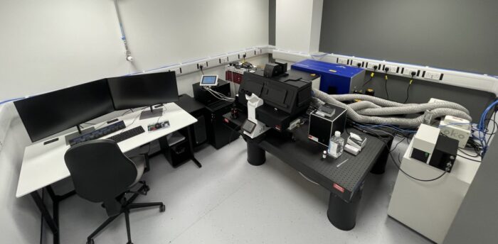
Hyperspectral BioImaging Facility

EPSRC funded Strategic Equipment: EP/Y01488X/1
The ability to visualise molecules, cells, tissues and other materials using optical microscopy is often limited by the researcher's ability to successfully label different components with a suitable dye, which can be inefficient and expensive, and also requires the researcher to know what they are looking for in advance of the experiment. In addition, imaging can be limited by how deep light can penetrate into dense samples such as tissues, reducing a researcher's ability to image complex structures such as tissues in 3D. Yet, single-molecule and live-cell imaging are recognised as invaluable tools in biophysics and bioengineering for many applications such as drug discovery, disease pathology and mechanistic biology. In addition, understanding the molecular distribution and structure of materials is critical for applications in drug delivery, food science, and regenerative medicine and needs to be studied across multiple length scales, from molecular to millimetres.
The Stellaris 8 CRS Microscope will enable researchers to map how different molecular species are distributed in their samples by observing their unique molecular fingerprints. This type of imaging allows imaging without requiring the 'labelling' or 'tagging' of components with dye molecules, providing real-time, high-resolution imaging of complex systems. It is beneficial for studying the distribution of different molecular components in food products, polymeric materials, and cellular components of tissue length. The system uses methods capable of penetrating deep into multicellular systems, providing unrivalled 3D resolution in tissue imaging. Since every molecule has a unique vibrational signature, the Stellaris 8 will allow the monitoring of drug distribution and accumulation in real-time, helping develop better models for studying disease and new methods for treating disease.
At the University of Leeds and beyond, a Stellaris 8 microscope will enable new science in areas of disease modelling and drug delivery; for example, understanding the biophysical and biochemical mechanisms of chemoresistance in cancer is critical to designing new therapeutics and understanding how new drugs interact with and distribute in model tissues facilitate drug discovery and improve patient outcomes in hard to treat cancers. In soft matter and biophysics, recent developments in hydrogel materials, lipid membranes and biomaterials in food science. In early detection and diagnosis, the advent of disease at the single-cell level is often accompanied by subtle changes in biochemistry and the spectral fingerprint. Detecting these early changes before advanced disease develops would ultimately save lives and reduce the health care costs to the nation.
The Stellaris 8 microscope will enable multimodal imaging and measurements across various biophysical research areas in a manner that is not currently possible, enhancing existing research and leading to new research and collaborations across multiple institutions.
About the microscope system:
Stellaris 8 FALCON platform: (click here for further info)
- White Light Laser (440-790nm) - pulsed
- 405nm Laser
- 2x high performance detectors
- 2x time resolved detectors for TauSense (click here for further info)
- Resonant scanner for video rate acquisition
- Standard confocal modalities: Z-stack, mosaics, time etc
- Lightning mode to 'stretch' the optical limit
- 2 photon and SHG fluorescence modes
Coherent Raman Scattering: (click here for further info)
- Fixed laser beam @ 1032nm
- Tunable laser beam @720-980nm
- SRS detector
- Epi-CARS detector
About the facility team:
Professor Stephen Evans - Head of School of Physics and Astronomy
Dr Julia Gala de Pablo - Lecturer in Biophysical Approaches for Healthcare
Dr Benjamin Johnson - Experimental Officer & HYBIFA Manager
How to apply for access:
Access will be free for an initial period, for proof of concept data etc. Further access may be chargeable. We will discuss this further with you following your application.
Please complete the access request form, by clicking here.
If you wish to submit additional information this can be added by replying to all, upon receipt of the confirmation email. We aim to respond, to all requests within 72 hours.
