
Tissue Culture and Analysis Facility
The Tissue Culture Lab within the MNP Research Group is designed to support advanced research at the intersection of physics, biology, and nanotechnology. This facility offers a controlled environment ideal for culturing and analysing a wide range of cell types, allowing researchers to investigate cellular behaviours, interactions, and responses at the nanoscale. By leveraging expertise in molecular and nanoscale physics, our lab facilitates studies that deepen our understanding of cellular mechanics, disease models, and tissue engineering.
Our lab is equipped with cutting-edge incubation, sterilization, and high-resolution microscopy systems, providing a robust platform for both foundational research and applied projects. Researchers can explore cellular responses to various physical and biochemical stimuli, with applications in areas such as drug discovery, regenerative medicine, and biomaterial development. The precision offered by our equipment enables the detailed analysis needed to observe cellular phenomena that are pivotal in health, environmental science, and biotechnology.
By bringing together the principles of physics with cell science, our tissue culture lab promotes interdisciplinary collaboration and innovation. Students and scientists gain hands-on experience with industry-standard technology, making our lab a valuable resource for training and development in cell culture techniques. Through this unique approach, our research group contributes to advancements that have broad-reaching implications for medicine, nanotechnology, and beyond.
The above image is a model made by Georgina Lee, where she is growing small blood vessel networks inside a tiny, flexible device made of PDMS (PolyDiMethylSiloxane). To create these blood vessel networks, she uses human cells from the lining of veins (specifically, from the umbilical cord, known as HUVECs-Human Umbilical Vein Endothelial Cells), and connective tissue cells called fibroblasts. By flowing cell culture medium through the device, she encourages the HUVECs to form connection with each other, resembling a blood vessel network. To make observation easier, the HUVECs are labelled with green fluorescent protein (GFP) and to assess network permeability, she introduces a red fluorescent solution (Texas Red Dextran), into the device to observe whether it remains confined within the vessel.
Cell Culture in Action
Discover our range of cutting-edge instruments, carefully chosen to support advanced research and detailed analysis. Each tool is selected to foster hands-on learning and inspire innovation.
HERACELL 240i and 150i Carbon Dioxide Incubators (Thermo Scientific)
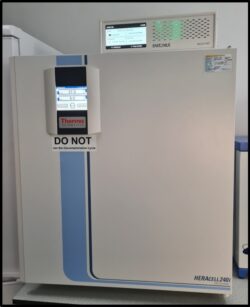
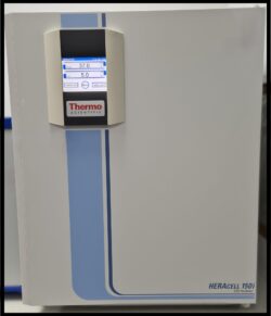
These CO2 incubators, 240i (combined with the Sartorius Incucyte SX1 Live-Cell Analysis System) and 150i, provide an optimal in vitro environment for precise cell culture. Equipped with thermal conductivity sensors, these incubators maintain consistent temperature and humidity levels, crucial for the stable growth of cells and prevention of contaminants.
The Heracell incubators are designed to support the delicate balance needed for cell growth, offering superior control over CO2 levels, temperature, and humidity. This creates a stable environment that fosters the growth, survival, and study of mammalian cells under ideal physiological conditions.
Currently, this setup at the MNP research group, is being utilized for research involving cancer mammalian cells, offering a reliable platform for long-term cell culture and live-cell imaging.
Incucyte (Sartorius SX1) - Live Cell Imaging and Analysis System

The Sartorius Incucyte SX1, housed inside the Heracell 240i CO2 incubator, is a cutting-edge live-cell imaging and analysis system that transforms cell-based research by providing real-time imaging and automated data analysis. It allows researchers to continuously monitor and quantify cellular behaviour over extended periods without disturbing the cells' natural environment. The system integrates effortlessly into standard incubators, making it a versatile and indispensable tool for laboratories conducting research in fields such as cell biology, immunology, oncology, and drug discovery.
The Incucyte SX1 captures high-quality images of live cells in real-time, enabling researchers to gain valuable insights into dynamic cellular processes such as proliferation, migration, and apoptosis. Its built-in algorithms facilitate automated data analysis, delivering accurate and quantifiable results while the experiments are ongoing. This capability eliminates the need for manual data collection, saving time and improving efficiency.
Designed to fit within standard tissue culture incubators, the Incucyte SX1 ensures that cells remain undisturbed in their optimal growth environment, preserving experimental integrity. Equipped with phase-contrast and fluorescence imaging capabilities, it supports a broad range of applications, from routine cell health monitoring to sophisticated, multi-parametric studies. Furthermore, its intuitive software interface simplifies experimental setup and data acquisition, making it accessible to researchers of all experience levels.
The versatility of the Sartorius Incucyte SX1 extends to its applications across diverse research areas. In cancer research, it enables the real-time tracking of tumor growth, invasion, and cell death. In immunology, the system facilitates the study of immune cell activation, migration, and interactions. Neuroscience researchers can use the Incucyte SX1 to observe neurite outgrowth and synaptic activity, while those in regenerative medicine can study stem cell differentiation and tissue formation. It is also a valuable tool for high-throughput drug discovery, supporting the screening of therapeutic compounds.
With its ability to capture continuous data, the Incucyte SX1 allows researchers to observe cellular processes as they occur, providing real-time insights. Its non-invasive imaging ensures that cells remain healthy throughout the experiments, while its automation reduces manual workload and accelerates research timelines. The system's scalability accommodates complex studies, such as multiplexed assays, enabling the simultaneous evaluation of multiple parameters.
Class II Tissue Culture Hood (Triple Red and Gelaire BSB BS5726)
 The Triple Red and Gelaire BSB BS5726 Class II MSC tissue culture hoods are highly efficient Biological Safety Cabinets (BSCs) designed to provide critical protection in laboratory environments. These BSCs serve as essential primary containment devices, safeguarding laboratory personnel from potential exposure to harmful biological agents by preventing splashes, aerosols, and contamination during tissue culture and other biological procedures.
Both hoods are engineered to create a sterile working environment, maintaining clean air through high-efficiency particulate air (HEPA) filtration. This ensures that airborne contaminants are effectively filtered out, protecting both the samples and the researchers. The airflow design of these Class II hoods also protects the work area from external contamination while simultaneously offering user protection, making them ideal for handling delicate biological materials, such as cell cultures, microorganisms, and hazardous biological agents.
In addition to protection, these BSCs promote optimal conditions for tissue culture by maintaining sterility and preventing cross-contamination, ensuring the integrity of experiments and research. This makes them indispensable tools for laboratories focusing on microbiology, virology, and cell culture work.
Microscope (VWR)
 The microscope with a mounted screen offers a powerful and user-friendly platform for precise imaging and analysis in various laboratory applications. This advanced microscope integrates a high-resolution screen, allowing for real-time viewing of specimens, which enhances collaboration and ease of use during microscopy tasks. The mounted screen (digital display capabilities – can capture, store, share images) eliminates the need for traditional eyepieces, offering a more versatile, comfortable and ergonomic experience for users (biological research, pathology, and medical diagnostics).
This setup is ideal for teaching, research, and clinical diagnostics, as it allows multiple viewers to observe specimens simultaneously, making it a great tool for group work and presentations. The microscope provides sharp, clear images with excellent contrast, making it suitable for a wide range of applications, including cell culture examination, tissue analysis, and microorganism identification.
Cell Counter (Countess 3)
 The Cell Counter is a highly efficient and easy-to-use automated cell counting device, designed to streamline the process of cell quantification in laboratories. With its advanced technology, this cell counter provides accurate and rapid cell counts in just a few seconds, eliminating the variability and time-consuming nature of manual counting under a microscope.
Equipped with a bright, high-resolution touchscreen interface, the Countess 3 allows for quick setup and operation, making it ideal for both novice and experienced users. The system is capable of counting live, dead, and total cells, as well as calculating cell viability, which is essential for applications such as cell culture, viability assays, and cell-based research.
In addition to its core function of counting, the Countess 3 can also analyse cell size and provide detailed insights into cell health and morphology. This makes it a versatile tool not only for routine cell culture work but also for more advanced research in fields like immunology, cancer research, and drug development.
The Countess 3's precision and ease of use significantly enhance lab productivity, providing consistent and reproducible results while minimizing errors and manual effort. It is an indispensable tool for laboratories focused on cell biology, tissue culture, and biomedical research.
Additional Instruments
The tissue culture lab is fully equipped with a comprehensive range of advanced instruments to support various cell culture and biological research activities. These instruments are essential for ensuring optimal conditions for cell growth, manipulation, and analysis, providing a reliable and controlled environment for scientific experimentation.
- Incubators (GS Biotech 48L, Grant JB Nova): These incubators maintain precise temperature, humidity, and CO2 levels to create ideal conditions for cell culture, ensuring consistent growth and viability of cells over extended periods.
- Sonicator (Ultrawave): The sonicator uses high-frequency sound waves to disrupt cells, homogenize samples, or dissolve substances, making it a versatile tool for sample preparation in cell culture applications.
- Mixers (SciQuip Vortex, Labnet Nutating Mixer): These mixers are designed to facilitate gentle or vigorous mixing of cell suspensions and reagents, ensuring uniform distribution and proper mixing of components.
- Hot Plate Stirrer (Stuart CD165): This device provides efficient heating and stirring for various laboratory tasks, from mixing reagents to dissolving substances in culture media.
- Syringe Pump (HA Harvard Apparatus PHD Ultra): This precision pump allows controlled delivery of small volumes of reagents or media, ensuring accurate and consistent dosing in experiments.
- Spinner (Sci Spin): The spinner supports cell culture by providing gentle agitation, promoting even distribution and aeration, which is particularly useful for growing suspension cultures.
These instruments, available for use in the tissue culture lab, offer a comprehensive suite of tools designed to facilitate cutting-edge research in cell biology, tissue engineering, and biotechnology. They ensure a controlled, sterile environment for culturing cells and performing essential analytical tasks, supporting a wide range of experimental protocols and research endeavours.